Heart Diagram Labeled Coronary Sinus : Angiographic imaging of left coronary sinus showing a ... : Coronary arteries supply oxygenated blood to the heart muscle, and cardiac veins drain away the blood once it has been deoxygenated.
Get link
Facebook
X
Pinterest
Email
Other Apps
Heart Diagram Labeled Coronary Sinus : Angiographic imaging of left coronary sinus showing a ... : Coronary arteries supply oxygenated blood to the heart muscle, and cardiac veins drain away the blood once it has been deoxygenated.. Venae cordisminimae (thebesian veins).coronary sinus it's. Coronary sinus (in coronary sulcus) (e). Even though the coronary sinus is major access. The aortic sinuses are small openings found within the aorta behind the left and right flaps of the aortic valve. Want to learn more about it?
After receiving oxygen from capillaries in the heart's muscle, the blood travels through cardiac veins, collects in the coronary sinus, and then flows into the atrium where the. The coronary arteries arise from the coronary sinuses immediately distal (superior) to the aortic valve and supply the myocardium with oxygenated blood. ▪ understand how to optimize imaging (ie how do i see a lesion in the lad better?) ▪ interpret coronary. 2 and 3 dimension transesophageal echocardiography images of the coronary sinus and middle cardiac vein in a patient with severe calcific aortic stenosis. Daniel nelson on january 1, 2019 1 comment 🤔.
Patente US8142493 - Method of heart valve repair ... from patentimages.storage.googleapis.com ▪ understand normal coronary anatomy ▪ understand different imaging views/projections. The heart, one of the most significant organs in the human body, is nothing but a muscular pump which pumps blood throughout the body. The human heart and its functions are truly fascinating. Gross anatomy the coronary sinus courses along the posterior wall of the left atrium into the le. The coronary sinus, the length of which varies from 15 to 65 mm, is found at the posterior part of the coronary sulcus on the diaphragmatic or posterior surface of the heart and is the principal collector of the venous blood of the heart. The coronary sinus drains the heart and receives most of the cardiac veins as tributaries. The right coronary artery arises from the anterior aortic sinus at the beginning of the ascending aorta. After receiving oxygen from capillaries in the heart's muscle, the blood travels through cardiac veins, collects in the coronary sinus, and then flows into the atrium where the.
The coronary arteries provide oxygenated blood to the heart.
Section through the atrial and ventricular septa viewed from behind. Right/left atria, right/left ventricles, pulmonary trunk, aorta, superior/inferior vena cavae, pulmonary veins, coronary sinus, right/left atrioventricular valves (tricuspid + bicuspid), chordae tendinae. The coronary arteries, namely the right coronary artery and the left coronary artery, arise in the root of the aorta and supply the myocardium and the rca usually supplies the heart's conduction system (sinus and av node) so that stenosis or occlusion of this vessel often leads to cardiac arrhythmias! 2 and 3 dimension transesophageal echocardiography images of the coronary sinus and middle cardiac vein in a patient with severe calcific aortic stenosis. The heart shown in fig.8.15 after removal of the floor of the coronary sinus. Anterior aspect of coronary circulation diagram. Part of the inlet septum interposes figure 8.16: Take home points cardiovascular medicine boards and clinical practice. Auricle, coronary sulcus, diaphragmatic surface, posterior interventricular sulcus, and base of heart. Cradle the heart from the oblique pericardial sinus, and with slight tension, sever the pulmonary vv. Anatomical structures were labeled according to the terminologia anatomica. Coronary circulation is the circulation of blood in the blood vessels that supply the heart muscle (myocardium). It is present in all mammals, including humans.
The heart shown in fig.8.15 after removal of the floor of the coronary sinus. The coronary sinus, the length of which varies from 15 to 65 mm, is found at the posterior part of the coronary sulcus on the diaphragmatic or posterior surface of the heart and is the principal collector of the venous blood of the heart. The right coronary artery arises from the anterior sinus of valsalva and courses through the right atrioventricular (av) groove between the right artium and right ventricle. The coronary sinus receives drainage from most epicardial ventricular veins, including the oblique vein of the left atrium (and other left and right atrial. The right coronary artery arises from the anterior aortic sinus at the beginning of the ascending aorta.
Normal Cardiac Anatomy - pedscards.com from sites.google.com The coronary sinus is a collection of veins joined together to form a large vessel that collects blood from the heart muscle (myocardium). The heart, one of the most significant organs in the human body, is nothing but a muscular pump which pumps blood throughout the body. The coronary sinus, the length of which varies from 15 to 65 mm, is found at the posterior part of the coronary sulcus on the diaphragmatic or posterior surface of the heart and is the principal collector of the venous blood of the heart. Coronary sinus (in coronary sulcus) (e). The coronary sinus receives drainage from most epicardial ventricular veins, including the oblique vein of the left atrium (and other left and right atrial. ▪ understand how to optimize imaging (ie how do i see a lesion in the lad better?) ▪ interpret coronary. Daniel nelson on january 1, 2019 1 comment 🤔. Venae cordisminimae (thebesian veins).coronary sinus it's.
Aortic valve and mitral valve location diagram.
The coronary sinus, the length of which varies from 15 to 65 mm, is found at the posterior part of the coronary sulcus on the diaphragmatic or posterior surface of the heart and is the principal collector of the venous blood of the heart. ▪ understand normal coronary anatomy ▪ understand different imaging views/projections. The heart, though small in size, performs highly significant functions that sustains human life. Venae cordisminimae (thebesian veins).coronary sinus it's. The human heart and its functions are truly fascinating. In this diagram, the heart has been removed and you are looking toward the posterior wall of the this sinus is a leftover from heart development in the embryo. Auricle, coronary sulcus, diaphragmatic surface, posterior interventricular sulcus, and base of heart. The right coronary artery arises from the anterior aortic sinus at the beginning of the ascending aorta. The heart is one of the hardest working organs in the body, and is responsible for pumping blood throughout the entire body. The right coronary artery arises from the anterior sinus of valsalva and courses through the right atrioventricular (av) groove between the right artium and right ventricle. Even though the coronary sinus is major access. The coronary sinus is a collection of veins joined together to form a large vessel that collects blood from the heart muscle (myocardium). The aortic sinuses are small openings found within the aorta behind the left and right flaps of the aortic valve.
Auricle, coronary sulcus, diaphragmatic surface, posterior interventricular sulcus, and base of heart. The human heart and its functions are truly fascinating. Aortic valve and mitral valve location diagram. Venae cordisminimae (thebesian veins).coronary sinus it's. Diagram and anatomy of the heart internal anatomy of the heart heart diagram:
Superficial Heart Flashcards - Cram.com from images.cram.com Heart structures and coronary circulation. Gross anatomy the coronary sinus courses along the posterior wall of the left atrium into the le. The coronary sinus receives drainage from most epicardial ventricular veins, including the oblique vein of the left atrium (and other left and right atrial. Venae cordisminimae (thebesian veins).coronary sinus it's. Coronary sinus (in coronary sulcus) (e). Sinus arrhythmia is discussed including the ecg criteria, cause and the treatment. ▪ understand how to optimize imaging (ie how do i see a lesion in the lad better?) ▪ interpret coronary. They branch and encircle the heart to cover its surface.
Anterior aspect of coronary circulation diagram.
Section through the atrial and ventricular septa viewed from behind. 2017 acc/aats/aha/ase/asnc/scai/scct/sts appropriate use criteria for coronary revascularization in patients with stable ischemic heart disease. This article describes the typical course, pattern and distribution of the coronary arteries and some of their respective named branches. The heart, though small in size, performs highly significant functions that sustains human life. In this diagram, the heart has been removed and you are looking toward the posterior wall of the this sinus is a leftover from heart development in the embryo. Sinus arrhythmia is discussed including the ecg criteria, cause and the treatment. Anterior aspect of coronary circulation diagram. The heart shown in fig.8.15 after removal of the floor of the coronary sinus. Auricle, coronary sulcus, diaphragmatic surface, posterior interventricular sulcus, and base of heart. After receiving oxygen from capillaries in the heart's muscle, the blood travels through cardiac veins, collects in the coronary sinus, and then flows into the atrium where the. Even though the coronary sinus is major access. Coronary sinus (in coronary sulcus) (e). The coronary arteries arise from the coronary sinuses immediately distal (superior) to the aortic valve and supply the myocardium with oxygenated blood.
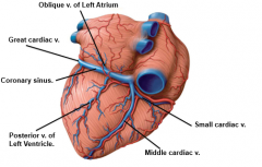
Locate the epicardium and features of the external, posteroinferior heart: coronary sinus labeled. It returns the majority of the blood supply for the left ventricle to the right atrium.


Comments
Post a Comment